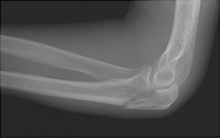

Besides, to facilitate adequate biplanar fluoroscopic imaging, the operating elbow must be extended and manually held outside the sterile surgical field, posing a risk of inadvertent loss of provisional fracture reduction tools. Despite the benefits of the supine position, it frequently requires a dedicated assistant to hold the elbow in the desired position throughout the surgery. In contrast, supine positioning is quick, provides easy access to the patient’s airway, allows for the management of concomitant injuries in a polytrauma setting, and facilitates gravity-assisted drainage of blood away from the operative field for better visualization. Prone or lateral decubitus positions are often considered, but they are time-consuming, limit the ability to manage patients in a polytrauma setting, and carry an increased risk of complications such as reduced stroke volume, cardiovascular collapse, rhabdomyolysis, and nerve palsy. When surgical fixation is deemed necessary, optimal patient positioning is critical for ease of surgical exposure, fracture reduction, and intraoperative imaging while avoiding complications. However, supine positioning minimizes setup time, limits the complexity of anesthetic monitoring, and accommodates management of patients with multiple injuries who cannot be positioned either lateral or prone.Olecranon fractures are common injuries accounting for 10% of all upper extremity fractures. Some surgeons prefer placing the patient either in a lateral or in a prone position. Often can be achieved with the elbow in 90 degrees of flexion, but most complex fractures require the freedom to freely flex and extend the elbow. Flexion of the elbow can be adjusted by varying the height of the support under the proximal forearm. 1, 2 Using nitinol staples to secure an olecranon osteotomy could improve this issue by providing. When an assistant is available, it is their job to support the arm. As much as 30 of olecranon osteotomy hardware has been removed as it irritated the patients. When an assistant is not available, the wrist may be secured with a sterile Kerlex (Kendall Healthcare Products, Mansfield, MA) and attached to a weight on the patient’s contralateral side. Surgery is performed with the forearm placed across the chest, with a supportive bolster placed beneath the proximal forearm ( Fig. Olecranon fracture is one of the most common elbow fractures, because the bone is not protected by soft tissue such as tendons, muscles or ligaments. Nondisplaced fractures can be treated with a short period of immobilization followed by gradually increasing range of motion. You can see the olecranon when you bend your arm. Fractures of the olecranon process of the ulna typically occur as a result of a motor-vehicle or motorcycle accident, a fall, or assault. It is covered by cartilage that allows it to move easily. To facilitate visualization and stability, the table is tilted obliquely toward the patient’s noninjured side. Olecranon fracture: The olecranon is the bony tip of the elbow and part of the ulna. The olecranon is the portion of the ulna (one of the bones in the forearm) that cups and fits tightly with the end of the humerus (upper arm bone). Initiate PROM of elbow 30-100 (greater extension is acceptable). A nonsterile tourniquet is applied to the upper arm. Exercises: Continue all exercises listed above. The patient is placed in a supine position.
LEFT OLECRANON FRACTURE SKIN
Because the olecranon is a subcutaneous bone, soft-tissue abrasions may require local skin care prior to internal fixation.Įither regional or general anesthesia can be utilized. For closed olecranon fractures, internal fixation is performed electively when the soft tissues permit.
LEFT OLECRANON FRACTURE SERIES
Low-velocity gunshot wounds without a neurovascular injury are treated with local wound care, antibiotic administration, and fracture stabilization if indicated. PMID: 27211227 DOI: 10.1016/j.injury.2016.04.015 Abstract Introduction: The aim of this study was to report the physical and functional outcomes after open reduction internal fixation of the olecranon in a large series of patients with region specific plating across multiple centres. If a vascular injury is present, exploration, repair, and external fixation should be performed urgently in collaboration with a vascular surgeon. In patients with highly comminuted fractures associated with grossly contaminated wounds, splinting or external fixation is preferred with sequential débridements followed by delayed Immediate internal fixation may be beneficial for Grade I and II open fractures, in patients who are hemodynamically stable. With open fractures, irrigation and débridement should be performed as soon as the patient’s condition and institutional resources permit. The timing of surgery for olecranon fracture fixation is determined by the status of the soft tissues.


 0 kommentar(er)
